Newer Development in Kidney Stone Removal

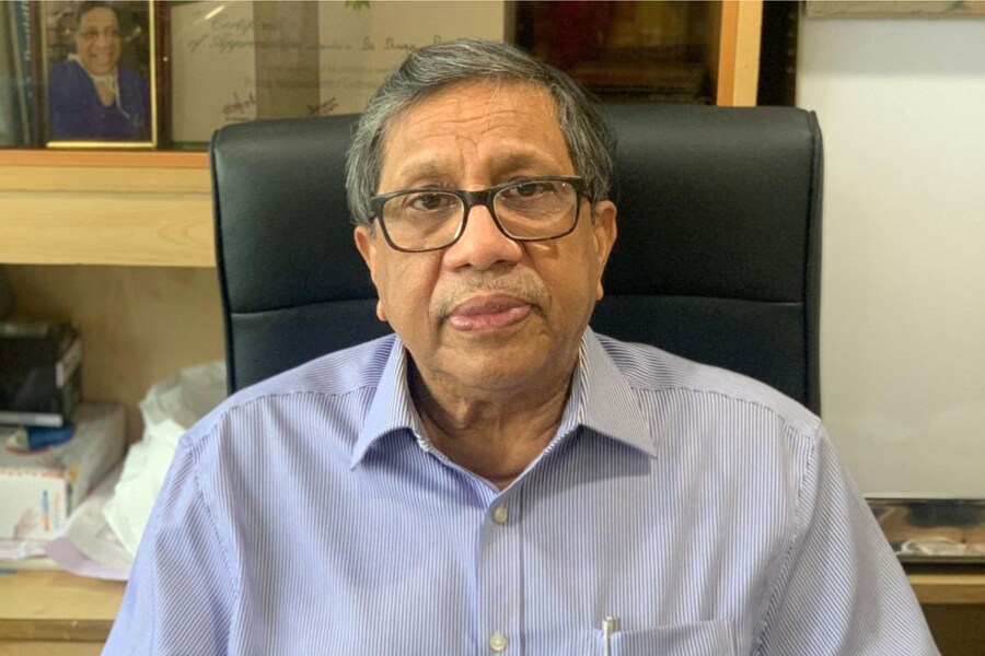
Dr. Shivaji Basu, MS, FRCS(Edin), FRCS(Eng), Director Urology, Fortis Hospital & Kidney Institute, Kolkata
BRAND CONNECT
Published: Nov 26, 2021 01:36:06 PM IST
Updated: Nov 26, 2021 05:37:57 PM IST

Introduction

Minimally invasive interventions for stone disease in the United States are mainly founded on 3 surgical procedures: extracorporeal shock wave lithotripsy, ureteroscopic lithotripsy, and percutaneous nephrolithotomy. With the advancement of technology, treatment has shifted toward less invasive strategies and away from open or laparoscopic surgery. The treatment chosen for a patient with stones is based on the stone and patient characteristics.

Each of the minimally invasive techniques uses an imaging source, either fluoroscopy or ultrasound, to localize the stone and an energy source to fragment the stone.

Multiple paths and technologic advances have been proposed in the field of urology and minimally invasive surgery to improve PCNL puncture. The most relevant contributions, however, have been provided by the application ofmedical imaging guidance, new surgical tools,motion tracking systems, robotics, and image processing and computer graphics.

Read More

With the advancements of technology, minimally invasive surgery for stone disease has been refined with less morbidity and increased rates of stone clearance.

Extracorporeal Shock Wave Lithotripsy

The underlying principle is to generate shock waves: piezoelectric, electrohydraulic, and electromagnetic. During propagation and transmission of a shock wave, energy is lost at interfaces with differing densities. Therefore, a coupling medium is necessary to minimize the dissipation of energy of a shock wave as it traverses the skin surface. The combination of several events during shock wave lithotripsy are thought to cause stone fragmentation: spallation, tear and shear forces, cavitation, quasi-static squeezing, dynamic squeezing, and stone fatigue.4 In summary, the successive shock wave pressure pulses result in direct forces that fragment the stones into smaller pieces. stones into smaller pieces. ESWL is the one truly noninvasive treatment for stones. The American Urological Association guidelines on the management of renal calculi support the use of ESWL for kidney stones.

Ureteroscopy (URS)

Fiberoptics allowed for endoscope size reduction and facilitated the development of steerable deflectable ureteroscopes. Technological advancements have resulted in smaller, actively deflectable, flexible endoscopes that are easier to manoeuvre into the upper urinary tract. These scopes combined with powerful and precise holmium laser lithotrites and a variety of endoscopic retrieval devices have further enhanced the successful outcome of ureteroscopy.

Percutaneous Nephrolithotomy

It is performed through a posterior calyx usually in the upper or lower pole depending on the stone location and proximity of adjacent organs. Once the access to the collecting system is obtained and the tract from the skin to the renal pelvis dilated using radiological assistance, energy sources are used to break the stone if it cannot be removed intact.

Pediatric

For pediatric patients with upper tract kidney stones, ESWL is an effective and safe treatment technique. The pediatric ureter is shorter, more elastic and distensible, thus permitting easier transmission of stone fragments and preventing ureteral impaction. Therefore, they require fewer shocks and lower energy for comparable stone clearance. Ureteroscopy with laser lithotripsy is also an effective way to treat stones in pediatric patients with similar success rates as in the adult population. PCNL offers good clearance rates with acceptable morbidity in the pediatric population.

Pregnancy

Acute obstructive nephrolithiasis during pregnancy can be very risky to both mother and fetus. These patients need to be managed with special care. Ureteral stent placement is a convenient method for temporizing ureteral obstruction from stone disease. Stent placement is usually done using minimal fluoroscopy or ultrasound. Ultrasound is used as a means of avoiding radiation altogether. If conservative management fails and there is no active infection, ureteroscopy and laser lithotripsy can be offered if clinically appropriate with an overall reported stone-free rate of 88.3%.

Recent ascents:

a)   Medical robotic for PCNL

The typical approach resorts to a surgical needle attached to a robotic arm that is driven—automatically or controlled by the surgeon—in a 3D or 2D imaging volume. In the 3D case, the imaging volume results from the acquisition of a 3D US or by overlapping multiple 2D US images, taking into account information from tracking systems, image processing algorithms, and computer graphics. The robotic arms, whose control is automatic, are also represented in volume imaging, using similar navigation systems.

b)    Computer-aided surgery for PCNL

It emphasizes the target structures of surgery and surrounding tissues in preoperative data, using image segmentation algorithms or computer graphics (direct volume or surface rendering). Then, the image processed data are superimposed and registered onto real-time intraoperative video (augmented reality) or static preoperative volume data (navigation software). The surgical tools are commonly updated using motion tracking systems, such as the Polaris infrared camera or the Aurora electromagnetic system, in real time and with millimetric precision.

Conclusion

Technological advances in the treatment of nephrolithiasis have led to safer and more efficient minimally invasive surgical treatments. Patient and stone characteristics are used to determine the most efficient treatment with the least risk. Because of the benefits of endoscopic surgery, future developments may focus on camera resolution, field of view and depth perception, tactile feedback, and improvements in mobility and handling of instruments.

The pages slugged ‘Brand Connect’ are equivalent to advertisements and are not written and produced by Forbes India journalists.

X